The kneecap, also known as the patella, plays an important role in knee movement and stability. It normally glides within a groove at the lower end of the thigh bone during bending and straightening. A patellar dislocation occurs when the kneecap moves out of this groove, usually towards the outer side of the knee. This can cause sudden pain, swelling and difficulty moving the leg. Some people experience a single episode following an injury, while others develop recurrent kneecap instability. Understanding the cause, associated damage and appropriate treatment is important for recovery and reducing the risk of another dislocation.
What Is Patellar Dislocation?
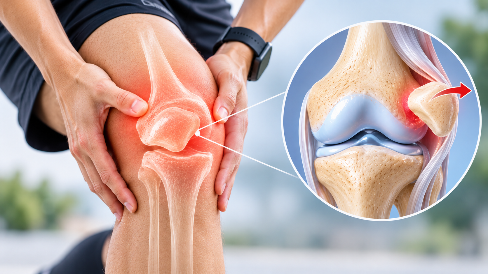
Patellar dislocation occurs when the kneecap completely moves out of its normal position within the femoral groove. A partial displacement, in which the kneecap temporarily moves away from its normal alignment and returns, is called patellar subluxation.
The patella is supported by surrounding muscles, ligaments and the shape of the femoral groove. The medial patellofemoral ligament (MPFL) helps prevent the kneecap from moving excessively towards the outside.
During a dislocation, supporting structures may stretch or tear. In some cases, cartilage or a small fragment of bone and cartilage may also become damaged.
A patellar dislocation is different from a general knee ligament injury or meniscus tear, although these conditions may sometimes occur together.
What Causes a Kneecap Dislocation?
Patellar dislocation commonly occurs during sports, sudden changes in direction, twisting movements or falls. A direct impact to the knee can also cause the kneecap to move out of position.
Certain anatomical factors may increase the risk of instability. These include a shallow femoral groove, patella alta (a relatively high-positioned kneecap), abnormal alignment of the lower limb and increased lateral movement of the patella.
Some individuals have naturally increased joint flexibility or a history of previous dislocations, which may contribute to recurrent instability.
A dislocation may occur during a relatively minor movement in someone with underlying anatomical predisposition. Identifying these factors helps guide treatment and decisions about recurrence prevention.
Symptoms of Patellar Dislocation
A sudden kneecap dislocation often causes immediate pain and difficulty moving the knee. The kneecap may visibly appear displaced towards the outer side, although it can sometimes return to its normal position spontaneously.
Common symptoms include sudden knee pain, swelling, tenderness around the kneecap, difficulty bending or straightening the knee and a feeling that the knee is unstable.
Some patients experience a sensation of the kneecap slipping or moving sideways during activity. This may be particularly noticeable while turning, climbing stairs or changing direction.
Rapid swelling may indicate bleeding inside the joint or associated cartilage injury. Persistent pain, recurrent instability or difficulty bearing weight should be assessed by a medical professional.
Symptoms alone cannot determine whether cartilage, bone or supporting ligaments have also been injured.
How Is Patellar Dislocation Diagnosed?
Diagnosis begins with a review of the injury, previous episodes of instability and the movements that caused symptoms. An orthopaedic examination assesses swelling, kneecap alignment, tenderness, movement and stability.
X-rays are commonly used to assess patellar alignment and identify fractures or bone abnormalities. They may also help evaluate anatomical features associated with recurrent dislocation.
MRI may be recommended to evaluate the medial patellofemoral ligament, cartilage, bone bruising and possible osteochondral injuries. It can provide important information when substantial swelling, persistent symptoms or associated damage is suspected.
In selected cases, additional imaging may be used to assess alignment or anatomical factors contributing to recurrent instability.
The findings from examination and imaging help determine whether treatment can begin conservatively or whether surgical assessment is appropriate.
First Aid and Initial Treatment for Patellar Dislocation
A suspected patellar dislocation should be evaluated promptly. If the kneecap remains displaced, urgent medical care is required for assessment and appropriate reduction.
Patients should avoid forcing the kneecap back into position themselves. Unnecessary movement may worsen associated injuries or cause additional pain.
Initial treatment may involve protecting the knee, applying cold packs with a protective cloth, elevating the leg and using appropriate support while awaiting medical evaluation.
A clinician may perform reduction when the kneecap remains dislocated. Following reduction, the knee is examined and imaging may be performed to identify fractures or associated cartilage injury.
The need for a brace, crutches or temporary restrictions depends on the injury and the treating clinician's assessment.
Non-Surgical Treatment for Patellar Dislocation
Many patients experiencing a first-time patellar dislocation without a significant associated osteochondral injury can be treated without surgery. The approach depends on knee stability, injury severity and individual risk factors.
Physiotherapy is an important component of treatment. It focuses on restoring knee movement, strengthening the quadriceps and hip muscles, improving balance and developing better control during functional activities.
Temporary bracing or other support may be recommended in selected cases, although prolonged immobilisation can contribute to stiffness and muscle weakness. The duration and type of support should be individualised.
Activity is gradually progressed as swelling decreases, movement improves and strength returns. Higher-impact sports and pivoting movements are usually reintroduced only after appropriate functional recovery.
Non-surgical treatment may be less successful in patients with repeated dislocations or significant structural abnormalities, making specialist reassessment important when instability continues.
When Is Patellar Dislocation Surgery Needed?
Surgery may be considered when the kneecap repeatedly dislocates, significant instability persists despite rehabilitation or an associated osteochondral injury requires treatment.
Medial patellofemoral ligament reconstruction is a procedure used to restore a key stabilising structure of the kneecap in selected patients with recurrent instability.
Some patients may require additional procedures to address contributing anatomical factors. The choice depends on the underlying alignment, patellar position, femoral groove shape and overall knee condition.
When a loose fragment of bone and cartilage is present inside the joint, surgical evaluation may be necessary to determine whether it can be repaired or whether another treatment is appropriate.
Surgery is not automatically required after every first-time dislocation. Decisions are based on associated injuries, recurrence risk, functional goals and clinical evaluation.
Recovery and Rehabilitation After Patellar Dislocation
Recovery depends on whether the injury is treated conservatively or surgically and whether cartilage, bone or ligament damage is present.
Early rehabilitation generally focuses on controlling swelling, restoring comfortable knee movement and gradually activating the quadriceps muscles.
Strengthening exercises progress to balance training, movement control and functional activities. Patients may eventually work on running, jumping and sport-specific movements when appropriate.
After surgery, rehabilitation may involve a brace, temporary weight-bearing restrictions and a structured physiotherapy programme. Progression is guided by healing, strength and functional milestones.
Returning to sport too early may increase the risk of recurrent instability. A clinician or physiotherapist should assess readiness before demanding activities are resumed.
Can Patellar Dislocation Happen Again?
A previous patellar dislocation can increase the likelihood of another episode, particularly in younger individuals or patients with underlying anatomical risk factors.
Recurrent dislocation may cause repeated damage to cartilage and supporting structures. Persistent instability can also reduce confidence during walking, exercise and sports.
Appropriate rehabilitation helps improve strength and movement control. Patients with recurrent episodes may benefit from a specialist assessment to identify anatomical factors that could require additional treatment.
Not every patient will experience another dislocation. The risk depends on the original injury, anatomy, age, activity level and treatment approach.
When Should You See an Orthopaedic Specialist?
Seek urgent medical evaluation if the kneecap appears displaced, the knee becomes severely swollen, you cannot bear weight or the injury causes significant pain and restricted movement.
An orthopaedic consultation is also recommended after a kneecap dislocation that has reduced spontaneously, particularly following a first episode.
Recurrent slipping, instability during sports or persistent discomfort after an earlier dislocation should not be ignored.
A specialist can assess associated cartilage damage, anatomical risk factors and the need for rehabilitation, further imaging or surgical treatment.
Conclusion
Patellar dislocation is an injury in which the kneecap moves out of its normal position, causing pain, swelling and instability. Some patients recover with appropriate rehabilitation, while others experience recurrent dislocations or associated cartilage injuries.
Accurate diagnosis, identification of anatomical risk factors and an individualised treatment plan are important for restoring knee function and reducing recurrence.
Early assessment by an orthopaedic specialist helps patients understand their treatment options and return to daily activities or sports safely.
Frequently asked questions
The kneecap moves out of its normal position within the femoral groove, usually towards the outer side of the knee. This can cause sudden pain, swelling and difficulty moving the leg.
Yes, sometimes the kneecap returns to its normal position spontaneously. However, the injury should still be evaluated because ligament, cartilage or bone damage may have occurred.
Not always. Many first-time dislocations without significant associated injuries can be managed with rehabilitation. Surgery may be considered for recurrent instability or selected associated injuries.
Recovery varies depending on injury severity and treatment. Some patients recover over several weeks with rehabilitation, while recurrent instability or surgery may require a longer recovery period.
Yes. Previous dislocation and certain anatomical factors may increase recurrence risk. Rehabilitation and specialist evaluation can help address instability and determine whether additional treatment is needed.
A dislocation may damage the cartilage on the kneecap or femoral groove and can sometimes produce an osteochondral fragment. Imaging may be recommended when associated injury is suspected.
Walking may be difficult or painful immediately after injury. Weight-bearing depends on the injury and associated damage, so medical assessment is important before resuming normal activity.
Dislocation refers to the kneecap moving completely out of its normal position, while subluxation is a partial displacement in which the kneecap does not fully leave the joint alignment.
Originally published on dranilraheja.com.