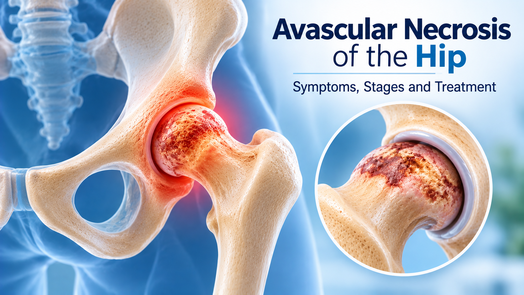
Avascular necrosis (AVN) of the hip, also called osteonecrosis of the femoral head, occurs when reduced blood supply damages bone tissue at the upper end of the thigh bone. As the affected bone weakens, it may gradually lose its structural strength and eventually collapse, damaging the hip joint. Treatment depends on the stage of disease, the extent of bone involvement and whether the femoral head has already collapsed. Selected early cases may be considered for hip preservation procedures, while advanced AVN with significant joint damage may require total hip replacement. The Direct Anterior Approach (DAA) is one surgical approach used for hip replacement in appropriately selected patients.
What Is Avascular Necrosis of the Hip?
Avascular necrosis is a condition in which bone tissue becomes damaged because its blood supply is reduced or interrupted. In the hip, it most commonly affects the femoral head, the rounded upper part of the thigh bone that fits into the hip socket.
Healthy bone requires an adequate blood supply to maintain its structure and repair itself. When blood flow is disrupted, bone cells may die and the affected region can gradually weaken.
As AVN progresses, the femoral head may develop a subchondral fracture or collapse. This can alter the shape of the joint surface and lead to secondary osteoarthritis.
AVN can affect one hip or both hips. The condition may progress at different rates depending on its cause, extent of involvement and stage at diagnosis.
What Causes Hip Avascular Necrosis?
Avascular necrosis may develop following a direct injury that disrupts blood vessels supplying the femoral head. Hip fractures and dislocations are important traumatic causes.
Long-term or high-dose corticosteroid exposure is a recognised risk factor for non-traumatic AVN. Steroids should never be stopped or changed without medical advice.
Excessive alcohol consumption is also associated with an increased risk of osteonecrosis. Certain medical conditions, including sickle cell disease and some blood or autoimmune disorders, may contribute to AVN.
In some patients, no definite cause is identified. Such cases may be described as idiopathic avascular necrosis.
Identifying potential risk factors and evaluating both hips when clinically appropriate may help guide assessment and monitoring.
Symptoms of Avascular Necrosis of the Hip
Early AVN may not produce noticeable symptoms. As the affected bone becomes weaker or structural changes develop, pain and movement-related discomfort may gradually appear.
Pain is commonly felt deep in the groin or front of the hip. Some patients also experience discomfort in the buttock or thigh, and pain may occasionally be perceived around the knee.
Symptoms may initially occur during walking, prolonged standing or exercise. As the condition progresses, pain may become more frequent, occur at rest or interfere with sleep.
Stiffness, reduced hip movement and difficulty bearing weight may develop in advanced stages. Patients may also experience reduced walking distance or difficulty performing everyday activities.
These symptoms are not specific to AVN and can occur with other hip conditions. Persistent hip or groin pain requires appropriate medical evaluation.
How Is Hip AVN Diagnosed?
Diagnosis begins with a review of symptoms, risk factors, previous injuries, medication exposure and relevant medical history. An orthopaedic examination assesses hip movement, pain, walking ability and functional limitations.
X-rays are commonly used to evaluate the hip joint and identify structural changes. However, early avascular necrosis may not be visible on standard radiographs.
MRI is particularly useful for detecting early AVN before significant changes appear on X-rays. It can help identify the affected region, assess the extent of bone involvement and evaluate the opposite hip when indicated.
CT may be used in selected cases to assess bone structure, subchondral fractures or femoral head collapse in greater detail.
Accurate staging helps determine whether the femoral head remains structurally intact and whether joint-preserving treatment may be appropriate.
Stages of Avascular Necrosis of the Hip
AVN is commonly classified into stages based on imaging findings, the extent of bone damage and the presence or absence of femoral head collapse. Different classification systems may describe these stages somewhat differently.
In early disease, MRI may show changes in the bone even when X-rays appear normal. Symptoms may be absent or limited during this phase.
As the condition progresses, structural abnormalities may become visible on imaging. A subchondral fracture can develop as the affected bone weakens.
Femoral head collapse represents a more advanced stage in which the rounded joint surface loses its normal shape. This can lead to increasing pain, restricted movement and secondary joint degeneration.
The stage of AVN is an important treatment consideration, but symptoms, lesion size, location, age and functional goals also influence management decisions.
Non-Surgical Treatment for Hip AVN
Non-surgical management may be considered in selected patients, particularly when symptoms are limited or the condition is being monitored. The aim is to manage discomfort, support mobility and address contributing risk factors.
Activity modification may involve reducing high-impact activities or prolonged weight-bearing that aggravates pain. In some situations, temporary use of walking aids may help reduce discomfort.
Pain-relief medication may be recommended when appropriate, after considering the patient's medical history and potential side effects.
Addressing contributing factors, including reviewing corticosteroid exposure with the prescribing doctor and reducing excessive alcohol consumption, may be important for overall risk management.
Non-surgical measures cannot reliably restore the blood supply or reverse established bone necrosis. Monitoring and follow-up imaging may be required to assess progression.
Hip Preservation Treatment for Early AVN
Hip preservation procedures aim to maintain the natural hip joint and delay or prevent femoral head collapse in selected patients. They are generally considered before advanced structural damage develops.
Core decompression is a procedure in which a channel is created within the affected femoral head to reduce pressure and support the biological response of the bone. It may be considered in selected early-stage cases.
Core decompression may sometimes be combined with other techniques or biological adjuncts. The potential benefits vary, and these approaches are not suitable for every patient.
The size and location of the necrotic area, stage of disease and condition of the surrounding bone influence the likelihood of benefit.
Hip preservation treatment does not guarantee that AVN will stop progressing. Continued monitoring and rehabilitation may be required after the procedure.
When Is Hip Replacement Needed for AVN?
Total hip replacement may be recommended when avascular necrosis has progressed to femoral head collapse or significant joint damage and symptoms substantially affect daily life.
The procedure involves replacing the damaged femoral head with an artificial component and preparing the hip socket to receive a prosthetic component. It aims to reduce pain and improve mobility and function.
Hip replacement may provide substantial improvement for appropriately selected patients with advanced AVN, but it involves surgical risks, rehabilitation and long-term considerations.
The decision depends on symptoms, structural damage, functional limitations, age, overall health and individual expectations.
Not every patient with AVN requires hip replacement. Early-stage disease may be evaluated for joint-preserving approaches, while some patients may initially be managed with monitoring and symptom control.
Direct Anterior Approach (DAA) for Hip Replacement in AVN
The Direct Anterior Approach is one surgical approach used to perform total hip replacement. It involves accessing the hip joint from the front of the hip through an interval between muscles, rather than routinely detaching major muscles from the bone.
In appropriately selected patients with advanced AVN, hip replacement through the Direct Anterior Approach may be considered when the femoral head has collapsed or the joint has developed significant damage.
The choice of surgical approach is based on several factors, including the patient's anatomy, body habitus, bone condition, previous surgeries, surgeon experience and the complexity of the hip problem.
DAA is not a treatment that reverses early AVN or restores the damaged femoral head. It is an approach for performing hip replacement when joint replacement is indicated.
The Direct Anterior Approach is not suitable for every patient, and other established approaches may be equally appropriate depending on individual circumstances and the surgeon's expertise.
Potential Benefits and Limitations of the Direct Anterior Approach
The Direct Anterior Approach uses an intermuscular and internervous interval, allowing access to the hip joint without routinely detaching certain major muscles from their bony attachments.
Some appropriately selected patients may experience less early muscle-related discomfort or quicker initial functional recovery. However, recovery varies considerably, and these potential advantages are not guaranteed.
The approach may allow early mobilisation after surgery when the patient's condition and surgical findings permit it. Rehabilitation and activity progression remain individualised.
DAA also has limitations. It involves a learning curve for surgeons and may carry specific risks, including wound complications, nerve irritation and difficulties related to exposure in certain patient anatomies.
No surgical approach is universally for every patient. The most appropriate approach is determined by patient factors, surgical complexity and the treating surgeon's experience.
How Is Hip Replacement Through the Direct Anterior Approach Performed?
Before surgery, the patient undergoes clinical assessment, imaging review and preoperative planning. The surgeon evaluates the extent of AVN, joint damage, bone anatomy and overall health to determine the appropriate procedure and approach.
During surgery, the patient is positioned according to the surgeon's technique. Access to the hip joint is obtained from the anterior side through the selected tissue interval.
The damaged femoral head is removed, and the hip socket is prepared to receive the artificial socket component. The femoral component is then implanted according to the surgical plan.
The surgeon assesses component positioning, hip stability, leg length and joint movement before completing the procedure.
The operation is performed under an appropriate anaesthetic plan. The exact technique, implants and surgical details may vary according to the patient's anatomy and the surgeon's practice.
Recovery After Direct Anterior Approach Hip Replacement
Recovery after hip replacement depends on the patient's health, extent of joint damage, surgical findings and rehabilitation progress. The Direct Anterior Approach does not guarantee a fixed or complication-free recovery timeline.
Many patients begin assisted mobilisation and walking soon after surgery when medically appropriate. A physiotherapist helps with walking practice, balance, exercises and safe movement.
Rehabilitation generally focuses on improving hip movement, strengthening surrounding muscles and gradually restoring independence in everyday activities.
Some patients may progress to routine activities within several weeks, while complete recovery and improvement in strength may take several months. Individual timelines vary.
Follow-up appointments help assess healing, implant position, pain and functional improvement. Patients should follow their surgeon's instructions regarding wound care, walking aids, driving, exercise and return to work.
Risks and Considerations of Hip Replacement Surgery
Hip replacement is an established procedure, but it carries potential complications. These may include infection, blood clots, dislocation, fracture, nerve or blood vessel injury, implant-related problems and differences in leg length.
Certain risks may vary according to patient factors, surgical approach and the complexity of the procedure.
The Direct Anterior Approach has specific considerations, including possible wound-related problems and nerve irritation in some patients. Risk assessment should be individualised.
Preoperative evaluation, appropriate surgical planning, rehabilitation and follow-up are important components of care.
Patients should discuss the expected benefits, limitations, alternatives and potential complications with their orthopaedic surgeon before deciding on surgery.
Can Avascular Necrosis of the Hip Be Prevented?
Not every case of AVN can be prevented, particularly when it is associated with trauma or certain medical conditions. However, some risk factors may be addressed.
Avoiding excessive alcohol consumption and using corticosteroids only as prescribed may help reduce certain risks. Patients taking long-term steroids should discuss concerns with their treating doctor rather than discontinuing medication independently.
People with recognised risk factors or a history of hip injury may require appropriate clinical monitoring. Persistent hip pain should be evaluated rather than ignored.
Early diagnosis is particularly important because treatment options may differ significantly before and after femoral head collapse.
When Should You See an Orthopaedic Specialist?
Consult an orthopaedic specialist if persistent groin or hip pain interferes with walking, exercise, standing or everyday activities.
Evaluation is especially important for individuals with previous hip trauma, prolonged corticosteroid exposure or medical conditions associated with osteonecrosis who develop new hip symptoms.
Seek prompt medical attention if severe pain develops suddenly, you cannot bear weight or symptoms follow a significant injury.
An orthopaedic assessment can help determine whether AVN is present, establish its stage and discuss appropriate treatment options, including hip preservation or replacement when indicated.
Conclusion
Avascular necrosis of the hip is a condition in which reduced blood supply damages the femoral head and may eventually cause joint collapse. Symptoms can develop gradually, and early disease may not be visible on X-rays.
MRI, appropriate staging and specialist assessment help identify the extent of bone involvement and guide treatment. Selected early cases may be considered for hip preservation procedures, while advanced disease with significant joint damage may require total hip replacement.
The Direct Anterior Approach is one option for performing hip replacement in appropriately selected patients with advanced AVN. Its suitability depends on individual anatomy, surgical complexity and the surgeon's expertise.
Timely evaluation and an individualised treatment plan help patients understand their options and make informed decisions about managing symptoms and restoring hip function.
Frequently asked questions
Avascular necrosis is a condition in which reduced blood supply damages bone tissue. In the hip, it commonly affects the femoral head and may eventually cause structural collapse and joint damage.
Early AVN may cause no symptoms. Some patients develop deep groin pain or discomfort during walking and weight-bearing activities, which may gradually become more persistent.
Yes. MRI is particularly useful for identifying early AVN when X-rays may still appear normal. It can also help assess the extent of bone involvement.
Selected early-stage cases may be considered for hip preservation procedures such as core decompression. Treatment suitability depends on the stage, lesion characteristics and condition of the femoral head.
The Direct Anterior Approach is a surgical technique for total hip replacement that accesses the joint from the front through an interval between muscles. It is one of several established approaches and is suitable for selected patients.
No. The choice depends on the patient's anatomy, body habitus, previous surgeries, bone condition, surgical complexity and the surgeon's experience. Other approaches may be equally appropriate.
Some selected patients may experience advantages in early recovery, but outcomes vary. The approach does not guarantee faster recovery or superior long-term results for every patient.
Hip replacement may be considered when femoral head collapse or advanced joint damage causes significant pain and functional limitations despite appropriate management.
Many patients begin assisted walking soon after surgery. Progress toward routine activities often occurs over several weeks, while strength and complete recovery may take several months depending on individual circumstances.
Yes. AVN can affect one or both hips. Depending on the clinical situation, a specialist may recommend evaluation of the opposite hip as well.
Originally published on dranilraheja.com.